Wednesday, October 24, 2012

Neuroetiquette and Neuroculture

Discover neuro-etiquette: fork and knife in action


Are neuroscientists taking jobs away from philosophers, sociologists and gender theorists?
"We need a neurocultural manifesto because the brain has been put forward by others as foundational for knowing about the self and social life, because neuroscientists are being asked to be the philosophers, sociologists and gender theorists of our era - they are being asked to do our jobs - and are responding with enthusiasm, and also because brain matter is mattering."

- from Neurocultures Manifesto by Victoria Pitts-Taylor.

In case you haven't realized yet, my job posting for a Tenure-Track Position in Neuroetiquette and Gender Theory was a spoof. The details were inspired by the highly unrealistic expectations of academia and by a disparate collection of neurowords.
  • Neuroetica -- I initially had a tough time decoding this word, translating its orthography to phonology and semantics. Neurorotica? Neuroetiquette? No, neuroetica is an Italian word from an article in Neuoethics that takes "a Look at the Development of the Italian Debate on Neuroethics." 

  • Neuroetiquette -- Much to my dismay, a writer at New York Etiquette Guide had already coined the word 'neuro-etiquette' in her blog post on neuroplasticity, learning to play the piano, and how to properly hold your knife and fork.

  • Neuroculture -- Etiquette is part of culture, of course. One encounters the word 'neuroculture' in many online discourses, from the tenets of the Neurocultures Manifesto to David Dobbs' blog Neuron Culture to the lifestyle marketing claim that Neuro Gasm Is Part Of The New Neuro Culture.

I think the neuro-panic among social scientists is overblown. How many philosophers, sociologists, and gender theorists are unemployed because their respective departments have decided to hire neuroscientists instead? How many developmental neurobiologists have applied for this Instructor of Philosophy position at Rochester Community and Technical College? Will a cognitive neuroscientst be able to teach transnational feminism or postcolonial feminism, queer theory, and critical race theory in the Women's and Gender Studies Program at Illinois State University?


Are we living in a neuroculture?

Daniel Buchman and David Dobbs asked that question two years ago. Their answers were "yes" and "of course we do!" More recently, a conference on Culture, Mind, and Brain: Emerging Concepts, Methods, Applications was held at UCLA:
The aim of this 2-day conference is to highlight emerging concepts, methodologies and applications in the study of culture, mind, and brain, with particular attention to: (1) cutting-edge neuroscience research that is successfully incorporating culture and the social world; (2) the context in which methods are used as well as the tacit assumptions that shape research questions; and (3) the kinds and quality of collaborations that can advance interdisciplinary research training. 
To find out what happened, you can check out the Conference Blog, Neuroanthropology, and Somatosphere. Ultimately, it sounds like we can all get along.

All sarcasm aside, I am in favor of multidisciplinary research. And I strongly endorse critical thinking about neuroscience. However, some self-appointed pontificators want to strip the brain of any power over human thought. At those times, it's good to see a defence of cognitive neuroscience. I'm starting a backlash against the anti-neuro backlash. After nearly 7 years of critical neuroblogging, it might be time for a change: The Neurocomplimenter.


But I never tire of highlighting those neuro-analogies that go over the top...
Discover neuro-etiquette: fork and knife in action

by Lyudmila Bloch, Etiquette Expert New York City

From the frontiers of neuroscience research, we know that our brain can change, reorganize, adapt, learn, and reprogram itself to a new “wiring” regardless of our age, previous experience, or current challenges.

A revolutionary discovery in neuroscience, called neuroplasticity, has confirmed that our brain is not a fixed, hardwired machine but rather vital and tirelessly evolving organ in our body. Experiments and clinical trials over the past two decades, conducted by the best minds in neuroscience, have discovered that our amazing brain, with proper rewiring and targeted conditioning, can master the most difficult of tasks at any age. Astonishing progress in overall functioning and new- skills acquisition show this master organ to be nothing short of, well, miraculous!

Leading behavioral psychologists and scientists have been collaborating, trying to understand the process of how a human brain learns and how it acquires new skills.
. . .

Any new skill or task we try to learn, including the proper use of dining utensils – so essential to our dining etiquette -- will entail the same kind of diligence in practicing, over and over, a “balancing exercise” -- holding your fork and knife correctly.

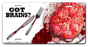
Got Brains? License Plate

Tenure-Track Position in Neuroetiquette and Gender Theory

Department of Critical Socioneurobiology.

Pending approval of departmental funds, the North Dakota School for Social Research is seeking outstanding candidates for its newly developed Interdisciplinary Program in Architecture, Kitchen Design, Sociology of Gender Roles, and Neuroimaging. State-of-the-art Siemens MAGNETOM 7T MRI and 306-channel planar dc-SQUID Neuromag Vectorview MEG facilities available. Start-up funds of $50K provided. Requirement to teach 3 classes per semester, including Statistics, Introduction to Celebrity Chefs, and Advanced Techniques in Optogenetics. The successful Assistant Professor candidate will be expected to obtain NEA funding, publish in high-impact science journals, give a Top 10 TED talk, and negotiate a major book deal before receiving tenure. Experience as a nationally syndicated advice columnist preferred.

Send CV, design portfolio, writing samples, research manifesto, and 10 letters of recommendation to: Chair of Search Committee, Department of Critical Socioneurobiology, North Dakota School for Social Research. Address inquiries to: neuroetiquette_and_gender_theory@ndsfsr.edu.

NDSFSR is an Equal Opportunity Employer.

Sunday, October 21, 2012

Savoir Faire or Savant?

Is amygdala volume correlated with social network size or with special talents in autism spectrum disorders? Or both??


The amygdala is a subcortical structure located within the medial temporal lobes. It consists of a number of different nuclei, or collections of neurons delineated by commonalities in morphology and connectivity. The amygdala is best known for major roles in fear conditioning (Paré et al., 2004) and responding to emotional stimuli more generally (Phelps & LeDoux, 2005), but its functions extend beyond that.

A presentation at the recent Society for Neuroscience meeting in New Orleans reported on MRI data obtained from Dr. Temple Grandin, the famous and talented Professor of Animal Science at Colorado State (Cooperrider et al., 2012):
Dr. Temple Grandin: A neuropsychological and multimodal neuroimaging case study of a savant with autism

. . .

Results: Dr. Grandin’s left lateral ventricle showed much more leftward volumetric asymmetry than controls. Her left cerebral white matter volume and bilateral entorhinal cortex thickness were much greater compared to controls. Her fusiform gyrus thickness was much less than the control mean. She had greater left lateral ventricle, intracranial, left cingulate, bilateral amygdala, and bilateral entorhinal cortex volumes. White matter microstructural differences were found for Dr. Grandin in multiple brain regions, including those known to relate to language function and facial information processing. ... The neuropsychological assessment indicated superior visuospatial and nonverbal reasoning abilities...

Virginia Hughes wrote a splendid summary of the study, which is not yet published in a peer-reviewed journal.

An earlier experiment was conducted with another very talented autistic savant (Corrigan et al., 2012):
The subject of this study is a 63-year-old, right-handed male with savant syndrome and a long-standing diagnosis of ASD.1 Institutionalized as a child, he has lived semi-independently as an adult, working for more than 30 years in dishwashing jobs. This individual is gifted with several special skills. One area of considerable talent is in music. He has perfect pitch and plays several musical instruments, of which his favorite is the accordion. He has substantial abilities with languages and can engage in basic conversations in twelve different languages. He also has remarkable abilities with sound imitation. His most exceptional ability, however, is in the area of art. ... He has become a highly regarded and accomplished graphic artist, whose works have been recognized through numerous exhibitions nationally as well as publication in a book. His medium is paper with pencil, marker, and crayon. His interest is in drawing collections, usually quite large, of items such as tools, birds, instruments, trains, flowers, and shoes, among many others. He takes a special interest in categorizing the physical world.

The volumes of his amygdala and caudate were both larger than values in the normative literature, and a strong right-sided asymmetry was seen for both structures (Corrigan et al., 2012). Hippocampal volumes did not differ from control values.

Although these two single-case studies were of exceptional people who may not be characteristic of the general ASD population, it was notable that both individuals had larger amygdalae than controls. Is this a surprising finding, in light of recent results on the correlations between amygdala volume and social network size in control participants? Surely the average neurotypical college student has a larger Social Network Index of offline contacts (Bickart et al, 2011) and more Facebook friends (Kanai et al., 2012) than the average person with ASD?
Grandin says, “the part of other people that has emotional relationships is not part of me” and she has neither married nor had children. ... She describes socializing with others as “boring” and has no interest in reading or watching entertainment about emotional issues or relationships.

In an earlier post on the Bickart article (More Friends on Facebook Does NOT Equal a Larger Amygdala), I noted that bigger is not always "better" (see Fig. 2 below):
One prominent example is the finding of larger amygdalae in children (and adults) with autism (Howard et al., 2000; Mosconi et al., 2009). However, the literature on this issue is variable (and voluminous)...2 More consistent are observations of increased amygdala volumes in generalized anxiety disorder (Etkin et al., 2009; Schienle et al., 2010). In rats, chronic stress causes hypertrophy (enhanced dendritic arborization) of pyramidal and stellate neurons in the basolateral nucleus of the amygdala (Vyas et al., 2002).


Modified from Fig. 2 (Howard et al., 2000). Volume estimation of the amygdala by the stereological point counting method. Section area estimation of posterior, middle, and anterior amygdala sections, using a regular array of test points. Section areas are increased in autism compared to controls.

A further interpretive conundrum is presented by the variety of conditions that are associated with increased amygdala volume: first-episode patients with nonschizophrenic psychoses, women high in harm avoidance, learning disabled adolescents at high risk of schizophrenia, adopted Romanian adolescents who experienced severe early institutional deprivation, and political conservatism.3
Most of those things are not especially fantastic for an active social life...

Autism is often considered as a disorder of microcircuitry and of long-range connections, so determining the structural and functional connectivity of the amygdala with other brain regions is crucial. One view holds that "underconnectivity" is a characteristic feature of the brains of those with autism, although recently this hypothesis has been called into question.

A new study from Bickert and colleagues (2012) followed up on their previous morphometric work and examined the functional connectivity of the amydala in relation to offline social network size in a group of 30 young adults (19 of whom had been in their previous experiment). Two separate groups of subjects, a "discovery sample" (n=89) and a "replication sample" (n=83) were scanned with fMRI at rest to establish the large-scale amygdala networks related to social cognition.

In brief, three networks were identified: (1) the ventrolateral amygdala and "perception" network (connected with lateral orbitofrontal cortex); (2) the medial amygdala and "affiliation" network (connected with ventromedial prefrontal cortex); and (3) the dorsal amygdala and "aversion" network (connected with dorsal anterior cingulate cortex).


Figure 1 (Bickert et al., 2012). Hypothetical topographic model of amygdala subregions and their affiliated large-scale networks subserving social cognition. A schematic of (a) the amygdala subregions in coronal view that we hypothesize are anchors for (b) three large-scale networks subserving processes important for social cognition. Ins, insula; SS, somatosensory operculum; dTP, dorsal temporal pole; cACC, caudal anterior cingulate cortex; rACC, rostral anterior cingulate cortex; sgACC, ubgenual anterior cingulate cortex; MTL, medial temporal lobe; FG, fusiform gyrus; vTP, ventral temporal pole; vlSt, ventrolateral striatum; vmSt, ventromedial striatum.


In the experimental sample (n=30), stronger intrinsic connectivity within the "perception" and "affiliation" networks was correlated with larger real-life social networks, but connectivity within the "aversion" network was not. A thorough evaluation of the methods used to establish these relationships is beyond the scope of this post, but an important question remains: can the current results inform the patterns of amygdala connectivity observed in participants with autism?


Footnotes

1 Although not specifically named, the participant was Gregory Blackstock, author of Blackstock's Collections: The Drawings of an Artistic Savant.


2 In fact, recent meta-analyses suggest that individuals with ASD may have smaller amygdala volumes than controls (Cauda et al., 2011; Via et al., 2011). On the other hand, the literature on early childhood hypertrophy of the amygdala in autism seems consistent (Mosconi et al., 2009). But another study in adolescents and adults with Asperger syndrome observed larger amygdala volumes than in controls (Murphy et al., 2012).

3 This study was published in a newspaper, not in a peer reviewed journal (see Left Wing vs. Right Wing Brains).


References

Bickart, K., Hollenbeck, M., Barrett, L., & Dickerson, B. (2012). Intrinsic Amygdala-Cortical Functional Connectivity Predicts Social Network Size in Humans. Journal of Neuroscience, 32 (42), 14729-14741. DOI: 10.1523/JNEUROSCI.1599-12.2012

Bickart KC, Wright CI, Dautoff RJ, Dickerson BC, Barrett LF. (2011). Amygdala volume and social network size in humans. Nat Neurosci. 14:163-4.

Cauda F, Geda E, Sacco K, D'Agata F, Duca S, Geminiani G, Keller R. (2011). Greymatter abnormality in autism spectrum disorder: an activation likelihoodestimation meta-analysis study. J Neurol Neurosurg Psychiatry 82:1304-13.

J.R. Cooperrider, E.D. Bigler, J.S. Anderson, S. Doran, C. Ennis, N. Adluru, A.L. Alexander, A.L. Froehlich, M.B.D. Prigge, J.E. Lainhart. Dr. Temple Grandin: A neuropsychological and multimodal neuroimaging case study of a savant with autism. Program No. 18.08. 2012 Neuroscience Meeting Planner. New Orleans, LA: Society for Neuroscience, 2012. Online.

Corrigan, N., Richards, T., Treffert, D., & Dager, S. (2012). Toward a better understanding of the savant brain, Comprehensive Psychiatry, 53 (6), 706-717. DOI: 10.1016/j.comppsych.2011.11.006

Etkin A, Prater KE, Schatzberg AF, Menon V, Greicius MD. (2009). Disrupted amygdalar subregion functional connectivity and evidence of a compensatory network in generalized anxiety disorder. Arch Gen Psychiatry 66:1361-72.

Howard MA, Cowell PE, Boucher J, Broks P, Mayes A, Farrant A, Roberts N. (2000). Convergent neuroanatomical and behavioural evidence of an amygdala hypothesis of autism. Neuroreport 11:2931-5.

Kanai R, Bahrami B, Roylance R, Rees G. (2012). Online social network size isreflected in human brain structure. Proc Biol Sci. 279:1327-34.

Mosconi MW, Cody-Hazlett H, Poe MD, Gerig G, Gimpel-Smith R, Piven J. (2009). Longitudinal study of amygdala volume and joint attention in 2- to 4-year-old children with autism. Arch Gen Psychiatry 66:509-16.

Murphy CM, Deeley Q, Daly EM, Ecker C, O'Brien FM, Hallahan B, Loth E, Toal F, Reed S, Hales S, Robertson DM, Craig MC, Mullins D, Barker GJ, Lavender T, Johnston P, Murphy KC, Murphy DG. (2012). Anatomy and aging of the amygdala andhippocampus in autism spectrum disorder: an in vivo magnetic resonance imagingstudy of Asperger syndrome. Autism Res. 5:3-12.

Paré D, Quirk GJ, Ledoux JE. (2004). New vistas on amygdala networks in conditioned fear. J Neurophysiol. 92:1-9.

Phelps EA, LeDoux JE. (2005). Contributions of the amygdala to emotion processing: from animal models to human behavior. Neuron 48:175-87.

Schienle A, Ebner F, Schäfer A. (2010). Localized gray matter volume abnormalities in generalized anxiety disorder. Eur Arch Psychiatry Clin Neurosci. Sep 5. [Epub ahead of print].

Via E, Radua J, Cardoner N, Happé F, Mataix-Cols D. (2011). Meta-analysis of graymatter abnormalities in autism spectrum disorder: should Asperger disorder besubsumed under a broader umbrella of autistic spectrum disorder? Arch Gen Psychiatry 68:409-18.

Vyas A, Mitra R, Shankaranarayana Rao BS, Chattarji S. (2002). Chronic stress induces contrasting patterns of dendritic remodeling in hippocampal and amygdaloid neurons. J Neurosci. 22:6810-8.

Saturday, October 13, 2012

Surrealistic Imaging Experiment #2


Scene from Le sang d'un poète (1930, Jean Cocteau) 1

"It is often said that The Blood of a Poet is a surrealist film. However, surrealism did not exist when I first thought of it."
-Jean Cocteau 2

In our second installment of Surrealistic Imaging Experiments, Marketing Professor Mohamed M. Mostafa of the Gulf University for Science and Technology in Kuwait was interested in how the brain processes Surrealistic imagery used in advertising (Mostafa, 2012). He approached the background to his topic and the fMRI study itself in a very different fashion from Silveira et al. (2012), who performed Surrealistic Imaging Experiment #1.

In that study, the authors hypothesized that humans cannot directly relate to surrealistic images because "percepts violating an expected percept cannot be integrated effortlessly into a frame of prior experiences or expectations and therefore also lack the potential for self-reference" (Silveira et al., 2012). Reduced activation in the precuneus to surrealistic relative to naturalistic pictures served as the major support for this contention. However, self-referential processing is also associated with other cortical midline structures (medial prefrontal and posterior cingulate cortices), which were not differentially activated here. Furthermore, there was no mention of top-down input from prefrontal cortex, networks for processing novelty, or lateral temporal regions associated with semantic processing. This seemed odd to me, as I expected that surrealistic paintings would require greater processing resources than realistic paintings.


Don't be lost in the weird world of investments.
Advertising Agency: Leo Burnett, São Paulo, Brazil


The Persistence of Memory in Advertising

In contrast to the restricted frame of reference presented above, Mostafa (2012) provides a historical background of Surrealism and summarizes the major themes and techniques used by iconic painters such as Salvador Dalí, René Magritte, and Giorgio de Chirico (who influenced the Surrealists). He extends the reach of surrealistic imagery to marketing and popular culture:
Today, the influence of Surrealism extends to cinema and advertising. Some Surreal advertisements create ambiguity by juxtaposing incongruous visual and verbal elements in a way that deliberately defies reason. It is not surprising that advertisers seek to create unique, unexpected and dreamlike images for use in promotions because advertisers often try to gain consumers' attention to fuel their fantasies and to induce them to view a product in a new light. For this reason, the casually Surrealist image has become, in particular, a stock-in-trade of the advertising industry (Macmillan 2000; Denny 2001).

Mostafa then segues into a review of neuromarketing and the brain imaging techniques used to probe the brains of consumers (fMRI, MEG, EEG). The aims of the study are articulated below:
...no previous studies have focused solely on investigating how consumers' brains process Surrealistic imagery in advertising. In this study, we aim to fill this void by investigating the neural correlates of Surrealistic imagery in advertising. More specifically, we aim to:

1.
identify cortical areas that selectively respond to Surrealistic imagery in advertising and
2.
test whether the elaboration and surprise hypotheses are supported within advertising context.3
Then, the specific hypotheses of the experiment are clearly elaborated after a review of the relevant literature:
H1: Surrealistic imagery in advertising will elicit more activation in brain areas associated with novelty detection such as the inferior frontal gyrus (IFG) and the anterior cingulate cortex (ACC).

H2: Surrealistic imagery in advertising will elicit more activation in brain areas associated with emotional responses such as the thalamus and the amygdala.

H3: Surrealistic imagery in advertising will elicit more activation in brain areas associated with episodic-memory retrieval such as the precuneus and the parietal cortex.

As for the methodological details, the participants were 18 right-handed English speakers in their 20s. None were experts in art history. The experimental task (passive picture viewing) used a 2 x 2 factorial design of (1) Surrealistic advertisements (novel, repeated) and (2) congruent advertisements (novel, repeated).

novel congruent    repeated congr   novel surrealistic   repeated surreal

Figure 1 (Mostafa, 2012). Stimuli and presentation sequence.


Thus, the author took into account both stimulus novelty and stimulus repetition (using a modified adaptation design), the latter to examine a form of priming in which a relative reduction in neural activity is observed when a stimulus is repeated. The stimulus set of Silveira et al. (2012) might have been better matched on size, luminance, color saturation, and spatial frequency parameters, but each of their 16 stimuli were repeated 3 times. There could have been differential priming effects for surrealistic vs. naturalistic images, but we don't know. On the other hand, the presentation parameters and number of stimuli in Mostafa's study weren't clear to me, either. 4

Briefly, the results confirmed the major hypotheses:
Surrealistic imagery elicited greater activation in several brain areas including the parietal cortex (BA 1, 2, 3), the precuneus (Brodmann area (BA) 7), the lateral parietal cortex (BA 39/40), the prefrontal cortex (BA 6/9), the IFG (BA 45/46), the ACC (BA 24), the insula (BA 13) and the amygdala. This result suggests that, compared to other types of advertisements, Surrealistic advertisements are processed in different functional brain pathways. This finding lends strong support to the elaboration and surprise hypotheses, which postulate that incongruous stimuli elicit increased processing that leads to more distinct and robust memory traces.

The novelty and distinctiveness of the Surrealistic images presented here in Surrealistic Imaging Experiment #2 required greater subcortical and cortical processing resources (including in the precuneus) than did the congruent images, in contrast to the detachment and alienation suggested by Surrealistic Imaging Experiment #1. How do we account for this discrepancy? What is the logical explanation?

"Our brains are dulled by this incurable mania for reducing the unknown to the known, to the classifiable. The desire for analysis wins out over feeling. It results in lengthy statements whose persuasive force derives from their very strangeness, and only impress the reader by recourse to an abstract vocabulary, which is moreover quite ill-defined."

-from the Surrealist Manifesto, by André Breton (1924)


Don't be lost in the weird world of investments.
Advertising Agency: Leo Burnett, São Paulo, Brazil


Footnotes

1 From Blood of a Poet: spectator as spiritualist
The film is essentially an exploration into the creative process; a self-conscious exploration of the subconscious, even the unconscious. Cocteau states that he was not thinking while he made the film but allowed images and ideas to present themselves to him, almost like the improvisation of a Jazz musician. Cocteau repeatedly spoke in interviews of the mysterious other self within an artist, saying that a poet (and by this he means artist of any genre) is a medium for the strange force which exists within him. He doesn’t shape this force, but rather the force shapes him. “My relationship with the work was like that of a cabinet maker who puts together the pieces of a table whom the spiritualists, who make the table move, consult”
2 This sly comment from Cocteau is not true; André Breton wrote the Surrealist Manifesto in 1924.

3 On memory, bizarreness, elaboration and surprise:
Prior behavioral research has also investigated visual imagery mnemonics and Surrealism in advertising. This line of research suggests that Surrealistic advertisements are likely to be processed more extensively than non-Surreal advertisements. For example, O'Brien and Wolford (1982) suggested that bizarre images increase the distinctiveness of items. Houston, Childers, and Heckler (1987) found that pictorial information incongruent with prior expectation is more difficult to comprehend and stimulates more elaborate internal processing. Behavioral support for bizarreness effect is based on both the elaboration hypothesis (Merry 1980; Wollen and Cox 1981) and the surprise hypothesis (Hirshman, Whelley, and Palu 1989). The elaboration hypothesis suggests that incongruous stimuli elicit extra processing because such stimuli are more difficult to make sense within the context of expected semantic norms.
4 It doesn't seem like the contrasts presented in Table 1 and Figs. 5 and 6 were corrected for multiple comparisons, which is a problem. The Silveira et al. (2012) study was methodologically superior in this specific realm.


References

Mohamed M. Mostafa (2012). The persistence of memory: an fMRI investigation of the brain processing of Surrealistic imagery in advertising. Journal of Marketing Communications DOI: 10.1080/13527266.2011.653688

Silveira S, Graupmann V, Frey D, Blautzik J, Meindl T, Reiser M, Chen C, Wang Y, Bao Y, PöppeI E, Gutyrchik E (2012). Matching reality in the arts: self-referential neural processing of naturalistic compared to surrealistic images. Perception 41:569-76.


Absurdly low consumption. The Polo BlueMotion.
Advertising Agency: DDB, Berlin, Germany

Tuesday, October 9, 2012

Surrealistic Imaging Experiment #1


"The case against the realist position needs to be considered, after considering the materialist position. The latter, more poetic however than the former, admittedly implies on the part of a Man, a monstrous pride, but not a new and more complete degeneration. It should be seen, above all, as a welcome reaction against certain ridiculous spiritualist tendencies. Ultimately, it is not incompatible with a certain nobility of thought.

The realistic position, in contrast, inspired by positivism, from Thomas Aquinas to Anatole France, appears to me to be totally hostile to all intellectual and moral progress. It horrifies me, since it arises from mediocrity, hatred and dull conceit. It is what engenders all the ridiculous books, and insulting plays of our day. It feeds on newspaper articles, and holds back science and art, while applying itself to flattering the lowest tastes of its readers; clarity bordering on stupidity, the life lived by dogs."

-from the Surrealist Manifesto, by André Breton (1924)

Surrealism was a philosophical movement in art and literature that embraced the unexpected, the impossible, the dream-like elements hidden from waking life. The founder of surrealism was French writer and poet André Breton, who was trained in medicine, psychiatry, and Freudian psychoanalysis. He defined surrealism as "Pure psychic automatism by means of which one intends to express, either verbally, or in writing, or in any other manner, the actual functioning of thought. Dictated by thought, in the absence of any control exercised by reason, free of any aesthetic or moral concern." Breton clearly had no use for the realist position, which he viewed as a pox on society. Eighty-eight years later, he might find it ironic that surrealism itself is the subject of positivist thought and scientific investigation.


Does the brain process surrealistic art in a different fashion than realistic art? 

Why yes, one would expect that to be the case. Undaunted, Silveira and colleagues (2012) asked this question in a recent fMRI study. I would predict that surrealist art is more computationally intensive for the Bayesian brain, so there would be greater activation in regions associated with top-down visual processing in an attempt to construct a coherent meaning from an unusual image.1 There was a precedent for this position in a study that compared the activations produced while viewing possible and impossible objects (Wu et al., 2012). Briefly, regions in both the dorsal and ventral visual streams showed greater activation for impossible than for possible objects.2


The authors of the current study adopted a different perspective (so to speak):
While naturalistic paintings present the world in a habitual way, surrealistic paintings violate the expected frame of reference. Surrealistic paintings are characterised by presenting mainly recognisable objects but in constellations that do not exist in real life or that are impossible from a physical point of view. In presenting impossible scenes, these artworks prevent an effortless processing of information to come to a meaningful interpretation of the visual world. ... We hypothesised that percepts violating an expected percept cannot be integrated effortlessly into a frame of prior experiences or expectations and therefore also lack the potential for self-reference. As it has been assumed that cortical midline structures refer to such self-representations (Han and Northoff 2008), we expected naturalistic and surrealistic paintings to have different activations in these areas.

The major prediction was that surrealistic images cannot engender self-referential processing, so differences in cortical midline structures (medial prefrontal and posterior cingulate cortices) were expected, with no further mention of dorsal/ventral stream visual regions, top-down input from prefrontal cortex, networks for processing novelty, or lateral temporal regions associated with semantic processing.


Figure S2 (Silveira et al., 2012). Reproductions of naturalistic and surrealistic artworks. N = naturalistic, S = surrealistic. N1: Edward Hopper “Sunday”. N2: Edward Hopper “Morning Sun”. N3: Eric Fischl “Japanese Bath”. N4: Francisco de Goya “La Cometa”. N5: Edward Hopper “Chop Suey”. N6: Francisco de Goya “Niños con Mastines”. N7: Edward Hopper “Summer Evening”. N8: Edward Hopper “Cape Cod Morning”. S1: René Magritte “Son of Man”. S2: Vladimir Kush “Pearl”. S3: Salvador Dalí “My Wife Nude”. S4: Max Ernst “Elephant Celebes”. S5: Vladimir Kush “Walnut of Eden”. S6: Giorgio de Chirico “Hector and Andromache”. S7: René Magritte “Le Prêtre Marié”. S8: Vladimir Kush “Departure of the Winged Ship”.


The final stimulus set of 8 realistic and 8 surrealistic paintings is shown above. These were selected from a larger set of images rated on valence, arousal, and familiarity by a separate group of participants. Highly familiar pieces were excluded.3 The control stimuli were homogeneous color fields. All pictures were matched in size, luminance, color saturation, and spatial frequency parameters. Each picture was presented 3 times, and the task was to make a binary decision about their reaction to the image: “Are you touched by the painting?” The subjects were equally affected by the realistic and surrealistic paintings, but took longer to decide on the realistic ones.

The major neuroimaging result was obtained using a subtraction analysis to compare the BOLD response to naturalistic vs. surrealistic paintings. There was significantly greater activation to the naturalistic pictures bilaterally in the precuneus (Brodmann area/BA 7) and medial occipital cortex (BA 17, 18, 19) and in the right middle temporal gyrus. It seemed that no brain areas showed greater activity while viewing the surrealistic images. In fact, the surrealistic pictures resulted in deactivation of the precuneus, medial occipital, and temporal regions when compared to the color field control condition.


Figure 1 (modified from Silveira et al., 2012). Neurometabolic level of processing for naturalistic versus surrealistic images. Sagittal section. Note: Prec = precuneus, MOC = medial occipital cortex.


Given some of the hypothesized roles of the precuneus, the authors' interpretation is plausible:
As a part of the cortical midline structures, precuneus activity has been suspected to be associated with visuo-spatial imagery, episodic memory retrieval, and even of what has been referred to as the “self ” (Cavanna and Trimble 2006). Episodic memory retrieval is very likely to be associated with top–down processing (Sestieri et al 2010). However, surrealist paintings are not only unfamiliar but also disturb our sense of consistency and coherence and may therefore also hardly be related to our sense of the self. The higher activation of the precuneus in the naturalistic condition supports the hypothesis that percepts matching expectations and therefore confirming specific aspects of reality can be linked both to
prior experiences and innate programmes of the representation of the visual world...

What doesn't make sense to me is why there were no visual areas that showed greater activation to the surrealistic images, as in the impossible object study (Wu et al., 2012). Furthermore, it is unclear why the processing of such marked visual/semantic incongruity would not activate any kind of "novelty" circuit in dorsolateral prefrontal cortex, temporal parietal junction, and hippocampus, or semantic regions in the lateral temporal lobe. A very interesting electrophysiological study (using non-invasive ERP recordings, or event-related potentials) found a component at 390 msec post-stimulus that was related to mismatches between scenes and objects, as shown below (Ganis & Kutas, 2003). The authors speculated that this N390 scene congruity response "reflects the action of visual scene schemata stored in the anterior temporal lobe."


 modified from Fig. 1 of Ganis & Kutas


A green apple floating in front of someone's face (S1), or a ship's sails made out of butterflies (S8), might involve a similar violation of visual scene schemata.

"We are still living under the rule of logic, that, of course, is what I am driving at. But in our day, logical procedures are only applicable in solving problems of secondary interest. The absolute rationalism still in fashion only allows us to consider facts directly related to our own experience. The aims of logic, in contrast, escape us. Pointless to add that our very experience finds itself limited. It paces about in a cage from which it is more and more difficult to free it. It leans, it too, on immediate utility, and is guarded by common sense. Under the flag of civilisation, accompanied by the pretext of progress, we have managed to banish from the spirit everything that might rightly or wrongly be termed superstition, fancy, forbidding any kind of research into the truth which does not conform to accepted practice. It was by pure chance, it seems, that a part of our mental world, and to my mind the most important, with which we pretended to be no longer concerned, was recently brought back to light."

-from the Surrealist Manifesto, by André Breton (1924)


Footnotes

1 So once again, we have a challenge for Bayesian theorists and their "free energy principle" (see Free-energy minimization and the dark-room problem), which seeks to reduce uncertainty.

2 See That's Impossible! How the Brain Processes Impossible Objects for details of that study:
The paper started by reviewing the basic neuroanatomy of the visual system and its division into dorsal ("where") and ventral ("what") visual streams. Objects are primarily represented in the ventral stream, and the lateral occipital complex (LOC) is one area that seems to be specialized for object recognition. The authors predicted that impossible objects would be difficult for the LOC to process; therefore, additional regions would be recruited...

What were the results? As predicted, regions in both dorsal and ventral visual streams showed greater activation for impossible than for possible figures: right superior parietal in the former and right fusiform and inferior temporal gyri in the latter.


3 The Magrittes weren't familiar? Especially “Son of Man”? Really?


References

Ganis G, Kutas M. (2003). An electrophysiological study of scene effects on object identification. Cog Brain Res. 16:123-44.

Silveira S, Graupmann V, Frey D, Blautzik J, Meindl T, Reiser M, Chen C, Wang Y, Bao Y, PöppeI E, Gutyrchik E (2012). Matching reality in the arts: self-referential neural processing of naturalistic compared to surrealistic images. Perception, 41 (5), 569-76 PMID: 23025160

Wu, X., Li, W., Zhang, M., Qiu, J. (2012). The neural basis of impossible figures: Evidence from an fMRI study of the two-pronged trident. Neuroscience Letters 508:17-21.


Yves Tanguy, Indefinite Divisibility (1942)

Wednesday, October 3, 2012

Law and Order: Psychiatry Unit


Crime dramas on American television are known for loosely adapting actual news stories "ripped from the headlines" and calling them fiction. Law and Order: Special Victims Unit is especially known for this pattern. For instance, in one episode last year a much beloved basketball coach who runs a charitable foundation was found guilty of sexually abusing his young players over the course of many years.


In another episode, a well-known politician's long-kept secret is finally revealed after 13 years: the child he fathered with the family housekeeper.


I thought I would do SVU executive producer Dick Wolf a favor and present a unique psychiatric case study that can be adapted for the small screen.  Since it's unknown to the American viewing public (and most everyone else), it will seem like a more original idea.


Patient Presenting with Stockholm Syndrome Ultimately Diagnosed with Münchausen Syndrome and Dissociative Identity Disorder

{SPOILER ALERT: it ends badly.}

A troika of unusual and spectacular psychiatric disorders was observed in one Dutch patient, as reported by Spuijbroek et al. (2012):
A young female of indeterminate age called a crisis hotline several times in the course of a month, giving accounts of severe sexual abuse, kidnapping by her father, and membership in a sect in the recent past. She spoke by preference to male staff members, was difficult to understand, and used a high-pitched, childlike voice. She provided different names and addresses that proved to be wrong. “God,” “men and women,” and “good and evil” were regular topics...

After a few weeks, she expressed suicidal feelings, saying she wished to go to God, and she agreed to be admitted to a local psychiatric hospital. She left, however, after 1 day. One month later, she was hospitalized again, using a different family name. She stated that she was 19 years of age and claimed to have been severely abused by her father and other sect members. ... Her isolation from the outside world, the power imbalance, her sympathy for her abusing father, and her unwillingness to escape the life-threatening situation were consistent with the clinical picture of Stockholm syndrome.

In cases of Stockholm syndrome, a person held hostage or captive comes to strongly identify and bond with her captor. Here, the patient claimed she was kidnapped into a religious cult and sexually abused, yet she felt sympathy for her abusers (which included her own father). However, it soon became apparent that she was not telling the truth:
At first, the personal information provided by the patient could not be verified. After several weeks,  however, her identity was revealed by police detection. The patient turned out to be a 27-year-old woman who had been reported missing 3 weeks earlier by her family. The patient had been receiving mental health care since childhood; she also had financial debts and was at risk of losing her housing. She had wandered about for several years, presenting regularly at various hospitals with a variety of somatic and psychiatric symptoms. She also frequently contacted the police with very detailed and alarming stories that were impossible to verify...

The patient at first seemed unaffected by the disclosure of her identity, but after some days she showed concern. She was uncomfortable at the reunion with her parents. Gradually her behavior changed: she answered to her own name now, and she would speak with female staff members. A few weeks later, the patient said that she could not remember anything about the period when she was admitted to the hospital...

Thus, paradoxical elements of both Münchausen Syndrome (deliberately fabricating an illness) and a dissociative disorder (involuntary rupture of memory, awareness, identity and/or perception) were observed. The diagnosis of Stockholm Syndrome was withdrawn, and psychotic disorders were ruled out:
...The patient did not report hallucinations. While her stories were often improbable, they were never bizarre, and no cognitive impairment was found. Given the many moments of acuity and social responsiveness displayed by the patient, the picture was inconsistent with schizophrenia. The family reported that the patient had fabricated fantastical stories since she was a little girl. Fugue-like states with periods of unexplained peregrination or wandering may have occurred as well. Nevertheless, the patient was regularly able to contact others, continuing her telephone calls to mental health workers and other health care professionals, in a very consistent way. After her identity was disclosed, “Munchausen syndrome,” with imitation of both mental and somatic conditions, turned out to be the most likely description of the patient’s condition.

An additional examination of the patient was conducted by specialists from a regional psychological trauma center... their assessment revealed the following cluster of severe dissociative symptoms: derealization, fugue, depersonalization, amnesia, identity confusion, pseudoepileptic seizures, sleep problems, and self-damaging behavior. Although it was recognized that the patient was suffering from pseudologia fantastica [pathological lying], her dissociative symptoms were regarded as serious and authentic...

The court ordered the patient to be hospitalized for 6 months. During this time she was prescribed a series of medications (including antidepressants, antipsychotics, and benzodiazepines), none of which were effective. She became suicidal. Although her suicide attempts were characterized as "ambivalent", it was unfortunate that she ultimately succeeded while in hospital:
After several months, suicidal tendencies and a desire for euthanasia were a regular topic of conversation for the patient. Several times during her hospitalization, she demonstrated overt suicidal behavior: two medication overdoses, once walking on railroad tracks, and twice putting a plastic bag around her head. These attempts seemed ambivalent, since the chance of detection by staff members was relatively high. The patient was placed in seclusion because of heightened suicidal risk several times, leading to increased fear and tension within the therapeutic relationship. After 7 months, the patient suicided on the ward by suffocation with a plastic bag...

The clinical case discussion ends with COMMENTARY by David A. Kahn, MD [imagine a voice-over by Rod Serling]:
Why have I been haunted by this case report of a mysterious, unfortunate woman who was never truly knowable? The treating staff was ambivalent in its diagnoses of both Munchausen syndrome, which is the intentional feigning of illness, and DID [dissociative identity disorder], or the unintentional adoption of two or more personalities or identities. These appear to be in contradiction, ironically a most accurate reflection of the emotions evoked by the patient who appeared both manipulative but also helplessly unable to control herself and tell a true story. Who was she? Did she know or not know? Was she putting them on, or was she actually unsure of who she really was? Her final act of suicide was a forceful if equally ambiguous communication, as non-verbal as her others, but the authors suggested that it indicated both a growing attachment to the first treatment team that would be lost with transfer to a specialized trauma program, and terror or rage at feeling either found out, or held captive. The first explanation, loss that could not be expressed in words, supports dissociation—an unconscious reaction to immense pain. The second explanation, exposure and entrapment, might suggest antisocial traces more consistent with Munchausen syndrome.

The expository feature of post-episode commentary by a medical expert would distinguish Law and Order: Psychiatry Unit from other series in the franchise. Or the show could be taken in a more haunting and profound direction, which would be a clear departure from the usual police procedural. The Psychiatric Twilight Zone, perhaps?


Reference

Spuijbroek EJ, Blom N, Braam AW, & Kahn DA (2012). Stockholm syndrome manifestation of Munchausen: an eye-catching misnomer. Journal of psychiatric practice, 18 (4), 296-303. PMID: 22805905